Section outline
-
Tiia Drenkhan-Maaten
Estonian University of Life Sciences, Institute of Forestry and Engineering, Chair of Silviculture and Forest Ecology

-
-
Preferred name: Armillaria root rot
Authority: Elias Magnus Fries (1821)
Taxonomic position: Basidiomycota, Agaricomycetes, Agaricales, Physalacriaceae
Other scientific names: Armillaria (Fr.) Staude
Common names in English: The Honey fungus
-
-
-
Many Armillaria species are economically damaging pathogens with a broad host range, which cause disease in coniferous and deciduous tree species and shrubs. Most woody plants are probably susceptible to the disease to some extent, also young and some non-woody plants may be attacked. Age of the host may influence the disease and older trees can tolerate infections much better and they survive much longer with infection. Older trees often get butt rot if they are infected rather than extensive killing of sapwood, cambium, and phloem. With conifers, killing of young, vigorous trees is common especially in plantations.
-
-
-
Armillaria is a root-infecting basidiomycete genus comprising a large number of species with a worldwide distribution. Currently, more than 40 Armillaria/Desarmillaria species have been officially described and recognized, but studies of species richness based on DNA sequences in public databases suggest that 50–60 Armillaria species may occur. In many cases, species of Armillaria are distinct between the Northern and Southern Hemisphere.
-
-
-
The effect of Armillaria on living host start with growth loss, leading to root and stem rot. Other symptoms of Armillaria root diseases are rather more nonspecific, including reduction of shoot growth, changes in foliage characteristics, crown dieback, stress induced reproduction, basal stem indicators, white rot decay and death. However, some signs of infection are specific to Armillaria species like the distinctive mycelial fans, characteristic rhizomorphs (also evident with in vitro cultures) as well as the characteristic tawny toadstools. In young trees and shrubs, the entire foliage may turn brown or wilt and death may appear to be sudden. In older trees, death may follow a gradual dieback and may be preceded by the production of sparse foliage or by an abnormally heavy crop of cones or fruit. As with several other tree root diseases, honey fungus infection may lead to resinous or gummy exudations from the lower part of the stem.
Symptoms of Armillaria root disease
Signs of Armillaria root disease
Reduced height/diameter growth
Armillaria basidiocarps (mushrooms) on tree base
Chlorotic foliage
Mycelial fans
Slow loss of foliage
Rhizomorphs
Distress cones
Yellow wet/stringy decay, papery when dry
Slow crown decline
Zone lines
Abundant basal resin flow on conifers
Gummosis on hardwood trees
Sudden or slow death of tree
-
-
-
Historically, the identification of Armillaria was largely based on the morphology of the basidiocarp, and interfertility or mating compatibility. In recent decades, recognition and identification of Armillaria species has become increasingly reliant on DNA sequences, such as phylogenetic analyses representing multiple gene regions, which elucidate the evolutionary relationships among the species.
Armillaria species are difficult to distinguish morphologically. In most cases, they are recognizable as distinct species: (a) they are intersterile; (b) there are some differences among them in morphology and molecular-genetic characters; (c) there are differences in distribution, host range, and virulence.
To confirm Armillaria root disease, the root collar and lower bole of the tree must be examined for those signs specific to the fungus or by culturing it onto agar from the host tissues. As with other diseases, early diagnosis is vital for successful control. Conventional identification techniques involve isolation of the fungus mycelium present and culturing on agar but this takes a longer time than for many other fungi hence the cultures are very prone to contamination. With the advent of the modern diagnostic techniques based on enzyme-linked immunosorbent assay (ELISA) and molecular methods based on nucleic acid, however, rapid identification of the disease has become possible.
-
-
-
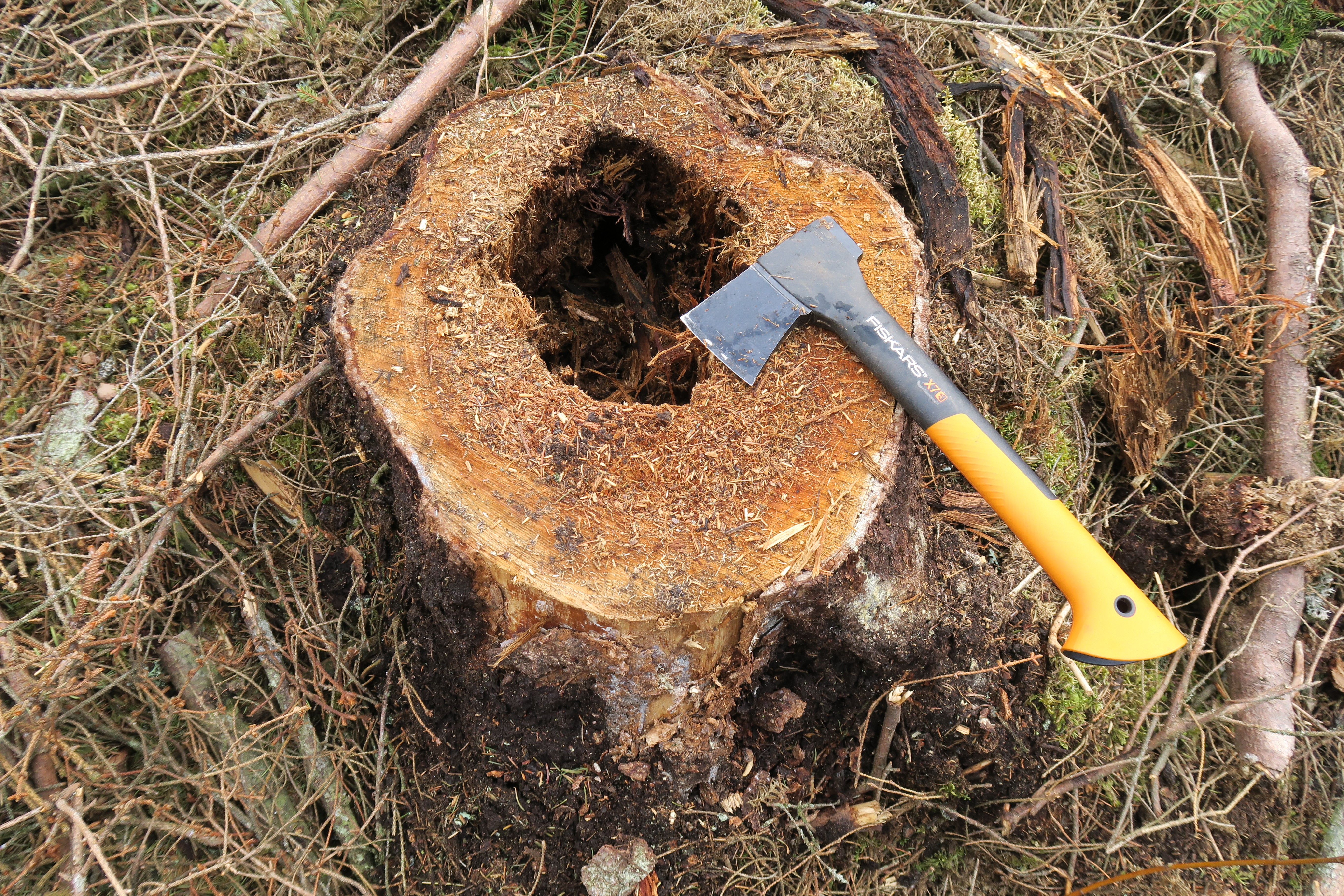
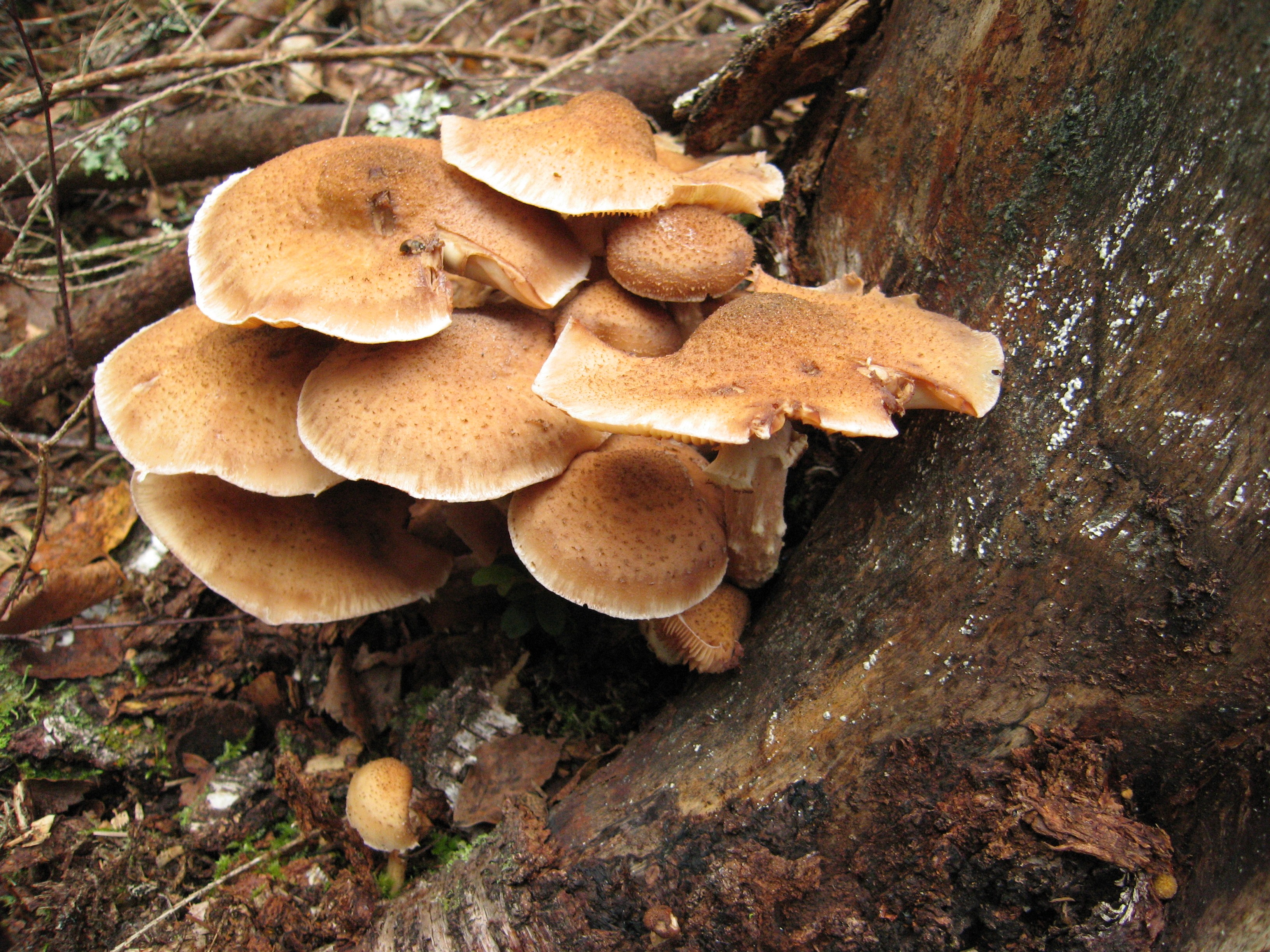
In addition to its pathogenic activity, the fungus lives saprophytically on tree stumps and other buried wood. Armillaria exists largely as a mass of microscopically fine threads (hyphae) growing inside the roots and butts of live trees, old stumps, fallen trees, and other woody debris. The fungus derives its nutrition mainly from the woody tissues, resulting in their gradual decay. Beneath the bark of roots and lower parts of the stem the hyphae may be so abundant that they form a clearly visible creamy-white fungal mass (mycelium). In some species, the rhizomorphs are quite tough and elastic but in others they are fragile. They are usually branched, though the pattern varies between species. Old rhizomorphs are almost black but younger ones are reddish brown with a white core and growing tip. Infections occur when growing rhizomorph tips contact and then penetrate roots or root collars of susceptible live plants. Spread may also take place without rhizomorphs via grafts and contacts between infected and uninfected roots. As well as infecting living trees, the fungus can colonise stumps and trees killed by other agents. All of these food sources serve as foci for further spread and infection. Honey fungus reproduces by means of microscopic spores produced in the toadstools. These spores are of no importance in the local spread of disease but provide the means by which the fungus can colonise more distant areas. Rhizomorphs are of limited value in the diagnosis of the disease: they may be difficult to find and can be confused with small tree-roots, whereas in A. mellea, which is probably the most pathogenic, rhizomorphs are usually fragile and hard to find.
-
-
-
In general, Armillaria infection tends to be more successful in susceptible hosts that are weakened, stressed, and/or maladapted to the current conditions on the site. After successful penetration through the bark, pathogenic Armillaria spp. produce mycelial fans in the vascular cambium beneath the bark of living trees, and these mycelial fans begin to degrade cellular components of the woody host. However, saprotrophic Armillaria spp. can also produce mycelial fans on dead trees or other woody materials. As infection progresses, roots and/or basal boles can be partially or fully girdled, and the tree continues to lose vigor, which also increases susceptibility to other pests and environmental stresses. After the host tissue dies, Armillaria is sustained by deriving nutrients obtained by colonization and degradation of dead wood and other organic matter. Armillaria is also capable of causing wood rot, such as heart rot, that can weaken the structural integrity of the basal bole and/or lateral roots. Trees with such wood rot represent hazard trees that are susceptible to structural failure and/ or wind throw, which can threaten life, limb, and property.
-
-
-
After becoming established in the roots, the vegetative mycelium of Armillaria develops a protective layer of thick-walled fungus cells, the pseudosclerotial envelope, about the body of the infected wood and the white mycelial fans in the cambium region. The rhizomorphs growing from the pseudosclerotium are also covered by this protective layer. The fungus may be acting as a secondary agent of damage in trees weakened by other means and can also grow saprophytically on trees killed by something else. When plants are stressed, for example, by drought or insect defoliation they become less resistant to the disease. As a result, infection of healthy roots more likely to occur but the pathogen may also be enabled to advance further in already infected roots. A major impediment in the chemical and biological control of Armillaria is the inability of the control agents to reach the site of inoculum inside wood in natural infections in sufficiently active state. The pathogen has also evolved highly sophisticated mechanisms of protection against outside deleterious effects. These include the production of antibiotics and the formation of pseudosclerotia. Armillaria spread typically occurs in the leaf-litter layer (duff) or underground via vegetative growth of rhizomorphs (dark, root-like mycelial structures) or mycelia (root-to-root contact). Rhizomorphs generally grow at a rate < 3 m per year, depending on the nutritional substrates, climate, and other environmental factors. In some situations, the radial growth of Armillaria results in an Armillaria root disease center or mortality center, but Armillaria often causes root disease that is more diffuse or scattered throughout the site.
- Short-range dissemination
Local spread is very important and is generally the dominant source of infection. The fungus can move by these means from an old root system, perhaps from a previous forest, to plants currently growing on the site. This leads to the appearance of disease in a previously uninfected population. The pathogen may survive for 50 years or more in stumps, it can wait until the new generation provides a large target for infection. The fungus can also move this way from a diseased tree to a neighboring healthy tree, leading to expanding areas of disease and mortality, usually called root disease centers. Root disease centers, as well as saprobic growth of the fungus, involve indeterminate growth through the forest. Resulting clones can cover many hectares, perhaps even miles, and be thousands of years old.
Mycelium can grow through direct root contacts and grafts with uninfected trees. Rhizomorphs can grow through soil to contact uninfected trees. Rhizomorphs are macroscopic, 1-5 mm diam., reddish brown to black, bundles of organized hyphae with an organized apically growing tip. Rhizomorphs grow through soil, produce branches, and look very much like roots (rhizomorph) or shoestrings (thus a common name for the disease, “shoestring root rot”). They use energy from a stump or killed tree to grow and infect a nearby tree. They can grow many meters through the soil.
- Long-range dissemination
Armillaria spp. is producing basidiospores, which are wind-dispersed to wreak death and destruction in new places, however, the spores don’t disperse very easily. There is indirect evidence that they do occasionally colonize stumps or wounds, especially in moist climates. Persistence in dead roots and stumps, the saprobic phase, may be dominant for some species that subsist by colonizing dead trees but rarely seem to kill them. Armillaria species may be abundant in the forest without a lot of obvious, damaging disease in some situations. Other species decay dead trees and stumps and build up energy to attack neighboring trees.
-
-
-
Armillaria and insects typically co-occur and exhibit strong interactions that are often difficult to interpret. Armillaria infection of spruce (Picea) can induce the production of volatile compounds that attract bark beetles, such as engraver beetles (Ips spp.). Because both, Armillaria and bark beetles tend to attack stressed trees, interactions among Armillaria and bark beetles are common, but temporal and causal relationships cannot always be definitively determined.
Another type of Armillaria-insect association includes defoliating insects, such as spongy moth (Lymantria dispar), eastern spruce budworm (Choristoneura fumiferana), maple webworm (Tetralopha asperatella), and saddled prominent caterpillar (Heterocampa guttivitta). In these interactions, insect defoliation is believed to predispose trees to Armillaria infections. Attack by root collar weevils (Hylobius spp.) has also been hypothesized to facilitate Armillaria infection by providing wounds that serve as points of entry for the pathogen. In many other cases, Armillaria and insects may simply co-occur without any direct interaction, while other Armillaria-insect interactions are yet to be examined.
-
-
-
Armillaria root disease occurs around the world in many places where woody plants grow. Depending on the environmental conditions, host plant, and host condition, different Armillaria species occur in different geographic regions, and each Armillaria species displays distinct ecological behaviors, ranging from a virulent primary pathogen, secondary pathogen, beneficial saprophyte, or beneficial mycorrhizal symbiont of orchids in Asia. Armillaria has some interesting, but disparate, features related to its ecological functions. For example, Armillaria is known for its bioluminescence, which is the subject of many hypotheses, but the ecological function of this property remains largely unverified. In eastern Asia, Armillaria spp. can form unique mycorrhizal relationships with achlorophyllous, mycoheterotrophic orchids, such as Galeola, Gastrodia, and Cyrtosia, which contain species that are important in traditional medicine. In other situations, Armillaria can participate in symbioses with other fungi, where it can serve as the host (e.g., Entoloma abortivum) or parasite (e.g., Wynnea).
-
-
-
- Physical removal of inoculum: by removing diseased trees and uprooting even neighbouring uninfected stumps. For example, trenches over a metre deep are dug to isolate the infected plants from healthy parts of a vineyard or a fruit orchard have been used to control spread of Armillaria root disease. Laying a plastic barrier in a trench and then backfilling it with the removed soil has been used for controlling the disease in kiwifruit orchards. Complete eradication of the fungus is also improbable and of doubtful value, and reinvasion by the fungus is possible. Soil disturbance may also stimulate fresh rhizomorph production, increasing the risk of disease to newly planted trees. Excessive removal of woody debris from a soil may be detrimental to antagonistic mycorrhizas.
- Stump removal: the most effective means of controlling the disease is to remove all sources of infection from the site. This normally means removing infected stumps and plants. The fungus may be present as rhizomorphs or minor root infections among trees and shrubs even where there is no sign of disease above ground. Therefore, if woody plants are removed for any reason, it is better to uproot them than to leave stumps in which the fungus could build up and become a greater threat to surrounding plants. If neither stump removal nor prevention of rhizomorph spread is possible, the only alternative is to leave stumps and infected trees where they are, accepting the inevitable losses, and to replant gradually with resistant species.
- Reducing root disease susceptibility: Because Armillaria seems to attack trees that are stressed due to climate maladaptation or other stress factors, a primary strategy for managing Armillaria root disease is to plant, select, or naturally regenerate trees that are well-adapted to the site and exhibit adaptation to a broad range of environments. Following a rotation with resistant species, the pathogen should have died out in the old root systems, permitting the reestablishment of susceptible species. In some situations, planted trees appear more susceptible to Armillaria root disease than naturally regenerated trees, especially if planted trees are not adapted to the site. Care must also be taken to avoid wounding trees that remain after thinning and use other methods to increase host vigor. In some horticultural situations, Armillaria-resistant rootstock may be available. Under horticultural conditions, the removal of soil surrounding the root crown may offer protection against Armillaria.
- Chemical treatments and fumigation: The most effective chemicals against A. mellea in vitro on 3% Malt Extract Agar were hexaconazole and flutriafol (both triazoles), fenpropidin (a piperidine), guazatine (a guanide) and phenylphenol (a phenolic compound) with Bray’s Emulsion as a standard.Volatile, toxic chemicals are placed in a hole drilled into an infested stump. The chemical kills the fungus or weakens it to the point other fungi can kill it. Even where stump extraction is effective at reducing Armillaria root disease, considerations should be given to (1) disturbances may allow new Armillaria inoculum to become established, (2) cost effectiveness, and (3) deleterious ecological consequences, such as reduced organic compounds/nutrients, soil erosion, and other long-term effects of mechanical disturbances. Still, chemicals for controlling Armillaria are either ineffective or are phytotoxic or have other environmental consequences, therefore is a need for biological control.
- Biological control: Antagonistic organisms might not be able to prevent Armillaria from becoming established in stumps, but they may restrict further stump colonisation and thus limit the available food base. An economical and environmentally friendly approach to biological control is the use of management practices to favor native, in situ biological control agents [e.g., Trichoderma spp. (fungi) and Pseudomonas (bacteria)] that are already present on the site, while discouraging the Armillaria pathogen. Among the most thoroughly studied fungal antagonists of Armillaria are Trichoderma species, common fungal hyperparasites abundant in the majority of soils. Mycophagous nematodes have also been implicated in the biological control of Armillaria. Studies in vitro have shown that ectomycorrhizal fungi can inhibit the growth of Armillaria. However, direct protection by mycorrhizas seems unlikely as the main infection sites for Armillaria are on larger roots rather than the fine roots where mycorrhizas develop.
Using management practices to favor native, in situ biological control agents does not require regulatory approval, and it focuses on the native biological control agents that are already adapted to the site. Methods, such as metagenomics and metabarcoding, can be used to determine fungi and bacteria present in the forest soil; however, knowledge of the microbiome and associated analyses are needed to determine which treatments (e.g., applications to adjust soil organic matter, N, and pH, and/or prescribed fire) favor the biological control agents and discourage the Armillaria pathogen.
Management of Armillaria root disease should focus on: (1) selecting or planting site-adapted tree/shrub species with less susceptibility to Armillaria, (2) using silvicultural methods to reduce tree stress, such as increasing tree spacing, (3) reducing inoculum of pathogenic Armillaria, (4) implementing management practices that favor natural biological control agents of Armillaria, (5) selecting or planting trees adapted to climate change and Armillaria root disease
-
-
-
1. Antonín V, Stewart JE, Medel Ortiz R, Kim M-S, Bonello P, Tomšovský M, Klopfenstein NB. 2021. Desarmillaria caespitosa, a North American vicariant of D. tabescens. Mycologia 113, 776–790. https://doi.org/10.1080/00275514.2021.1890969.
2. Baumgartner K. 2004. Root collar excavation for postinfection control of Armillaria root disease of grapevine. Plant Dis, 88, 1235–1240. https://doi.org/10.1094/PDIS.2004.88.11.1235.
3. Baumgartner K, Coetzee MPA, Hoffmeister D. 2011. Secrets of the subterranean pathosystem of Armillaria. Mol. Plant Pathol. 12, 515–534. https:// doi.org/10.1111/j.1364-3703.2010.00693.x.
4. Blodgett JT, Worrall JJ. 1992. Distributions and hosts of Armillaria species in New York. Plant Disease 76(2):166–169.
5. Burrill EA, Worrall JJ, Wargo PM, Stehman SV. 1999. Effects of defoliation and cutting in eastern oak forests on Armillaria spp. and a competitor, Megacollybia platyphylla. Canadian Journal of Forest Research 29(3):347–355 https://forestpathology.org/pdfs/burrill1999megacollybia.pdf.
6. Elías-Román RD, Calderón-Zavala G, Guzmán-Mendoza R, Vallejo-Pérez MR, Klopfenstein NB, Mora Aguilera JA. 2019. ‘Mondragon’: a clonal plum rootstock to enhance management of Armillaria root disease in peach orchards of Mexico. Crop Prot. 121, 89–95. https://doi.org/10.1016/j.cropro.2019.03.011.
7. Ferguson BA, Dreisbach TA, Parks CG, Filip GM, Schmitt CL. 2003. Coarse-scale population structure of pathogenic Armillaria species in a mixed-conifer forest in the Blue Mountains of northeast Oregon. Can. J. For. Res. 33, 612–623. https://doi.org/10.1139/X03-065.
8. Gregory SC, Rishbeth J, Shaw III CG. 1991. Pathogenicity and virulence. In: Armillaria Root Disease: Agricultural Handbook vol. 691, eds Shaw III CG, Kile GA, pp. 76–87. Washington, DC: USDA Forest Service.
9. Guo T, Wang HC, Xue WQ, Zhao J, Yang ZL. 2016. Phylogenetic analyses of Armillaria reveal at least 15 phylogenetic lineages in China, seven of which are associated with cultivated Gastrodia elata. PLoS One 11 (5). https://doi.org/10.1371/journal.pone.0154794, e0154794.
10. Heinzelmann R, Dutech C, Tsykun,T, Labbé F, Soularue J-P, Prospero S. 2019. Latest advances and future perspectives in Armillaria research. Can. J. Plant Pathol. 41, 1–23. https://doi.org/10.1080/07060661.2018.1558284.
11. Ibarra Caballero JR, Lalande BM, Hanna JW, Klopfenstein NB, Kim M-S, Stewart JE. 2022. Genomic comparisons of two Armillaria species with different ecological behaviors and their associated soil microbial communities. Microb. Ecol. https://doi.org/10.1007/s00248-022-01989-8.
12. Kedves O, Shahab D, Champramary S, Chen L, Indic B, Bóka B, Nagy VD, Vágvölgyi C, Kredics L, Sipos G. 2021. Epidemiology, biotic interactions, and biological control of Armillarioids in the Northern Hemisphere. Pathogens 10, 76. https://doi.org/10.3390/pathogens10010076.
13. Kim M-S, Heinzelmann R, Labbé F, Ota Y, Elías-Román RD, Pildain MB, Stewart JE, Woodward, S, Klopfenstein NB. 2022. Chapter 20 - Armillaria root diseases of diverse trees in wide-spread global regions. Forest Microbiology, Volume 2: Forest Tree Health, Volume 2: in Forest Microbiology, 361-378. https://doi.org/10.1016/B978-0-323-85042-1.00004-5
14. Klopfenstein NB, Stewart JE, Ota Y, Hanna JW, Richardson BA, Ross-Davis AL, Elías-Román RD, Korhonen K, Keča N, Iturritxa E, Alvarado-Rosales D, Solheim H, Brazee NJ, Łakomy P, Cleary MR, Hasegawa E, Kikuchi T, Garza-Ocañas F, Tsopelas P, Rigling D, Prospero S, Tsykun T, Bérubé JA, Stefani FOP, Jafarpour S, Antonín V, Tomšovský M, McDonald GI, Woodward S, Kim M-S. 2017. Insights into the phylogeny of Northern Hemisphere Armillaria: Neighbor-net and Bayesian analyses of translation elongation factor 1-α gene sequences. Mycologia 109, 75–91. https://doi.org/10.1080/00275514.2017.1286572.
15. Koch RA, Herr JR. 2021. Global distribution and richness of Armillaria species inferred from public databases and amplicon sequencing datasets. Front. Microbiol. 12. https://doi.org/10.3389/fmicb.2021.733159, 733159.
16. Koch RA, Wilson AW, Séné O, Henkel TW, Aime MC. 2017. Resolved phylogeny and biogeography of the root pathogen Armillaria and its gasteroid relative, Guyanagaster. BMC Evol. Biol. 17, 33. https://doi.org/10.1186/s12862-017-0877-3.
17. Korhonen K. 1978. Interfertility and clonal size in the Armillariella mellea complex. Karstenia 18, 31–42. https://doi.org/10.29203/ka.1978.135.
18. Kubiak K, Z˙ ółciak A, Damszel M, Lech P, Sierota Z. 2017. Armillaria Pathogenesis under Climate. Forests, 8(4), 100; https://doi.org/10.3390/f8040100
19. Labbé F, Lung-Escarmant B, Fievet V, Soularue J-P, Laurent C, Robin C, Dutech C. 2017. Variation in traits associated with parasitism and saprotrophism in a fungal root-rot pathogen invading intensive pine plantations. Fungal Ecol. 26, 99–108. https://doi.org/10.1016/j.funeco.2017.01.001.
20. McDonald GI. 2012. Is stumping a wise solution for the long-term: the problem of phenotype-environment mismatchin. In: Zeglen, S., Palacios, P. (Compilers), Proceedings of the 59th annual Western International Forest Disease Work Conference. 10–14 October 2011, Leavenworth, WA, USA. U.S. Department of Agriculture, Forest Service, Forest Health Protection, pp. 53–64.
21. Mihail JD. 2015. Bioluminescence patterns among North American Armillaria species. Fungal Biol. 119, 528–537. https://doi.org/10.1016/j. funbio.2015.02.004
22. Redfern DB, Filip GM. 1991. Inoculum and infection. In: Armillaria Root Disease: Agricultural Handbook vol. 691, eds Shaw III CG, Kile GA, pp. 48–61. Washington, D.C.: USDA Forest Service.
23. Rishbeth J. 1968. The growth rate of Armillaria mellea. Trans. Br. Mycol. Soc. 51, 575–586. https://doi.org/10.1016/S0007-1536(68)80027-0.
24. Schnabel G, Agudelo P, Henderson GW, Rollins P.A. 2012. Aboveground root collar excavation of peach trees for Armillaria root rot management. Plant Dis. 96, 681–686. https://doi.org/10.1094/PDIS-06-11-0493.
25. Sierota Z, Grodzki W. 2020. Picea abies–Armillaria–Ips: a strategy or coincidence? Forests 11, 1023. https://doi.org/10.3390/f11091023.
26. Smith ML, Bruhn JN, Anderson JB. 1992. The fungus Armillaria bulbosa is among the largest and oldest organisms. Nature 356, 428–431. https:// doi.org/10.1038/356428a0.
27. Stewart JE, Kim M-S, Lalande B, Klopfenstein NB. 2021. Pathobiome and microbial communities associated with forest tree root diseases. In: Asiegbu, F.O., Kovalchuk, A. (Eds.), Forest Microbiology -Tree Microbiome: Phyllosphere, Endosphere, and Rhizosphere. Vol. 1. Academic Press, Elsevier, Inc, London, UK, pp. 277–292, https://doi.org/10.1016/B978-0-12-822542-4.00004-8. Chapter 15.
28. Suetsugu K, Matsubayashi J, Tayasu I. 2020. Some mycoheterotrophic orchids depend on carbon from dead wood: novel evidence from a radiocarbon approach. New Phytol. 227, 1519–1529. https://doi.org/10.1111/nph.16409.
29. Wargo PM, Houston DR. 1974. Infection of defoliated sugar maple trees by Armillaria mellea. Phytopathology 64, 817–822. https://doi.org/10.1094/ PHYTO-64-817.
30. Warren GL, Singh P. 1970. Hylobius Weevils and Armillaria Root Rot in A Coniferous Plantation in Newfoundland, Nfld. Bi-Monthly Research Notes. 26 Canada Fisheries and Forestry, Canadian Forestry Service, St. John, NL, Canada, p. 55.
31. van der Kamp BJ. 1993. Rate of spread of Armillaria ostoyae in the central interior of British Columbia. Can. J. For. Res. 23, 1239–1241. https://doi. org/10.1139/x93-156.
-
-